4

Back to all posts

RenoVō® CASE STUDY: Superficial Digital Flexor Tendon (RH)

Angie Esselman, DVM, MS, DACVSMR
Rocking E Equine Sports Medicine and Rehabilitation | Stillwater, Oklahoma

Case Overview

I evaluated a 6-year-old western performance horse used for cutting that presented for lameness. The lameness was localized to the medial femorotibial joint using diagnostic analgesia. Based on this localization and clinical presentation, I proceeded with further imaging and diagnostic workup to characterize the underlying pathology. 

Presenting Signs & Functional Limitations

At presentation, the primary concern was performance-limiting lameness. The horse had been actively competing prior to the onset of clinical signs, and the lameness interfered with its ability to continue working in a cutting program.

No additional systemic abnormalities were noted. The issue appeared isolated to the affected stifle joint, with functional limitations consistent with intra-articular pathology impacting performance. 

Baseline Objective Assessment

After localizing pain to the medial femorotibial joint, I obtained radiographs, which were unremarkable. Given the persistence of lameness, I proceeded with ultrasonographic evaluation of the stifle.

Ultrasound identified a medial meniscal injury, which supported the decision to move forward with arthroscopic evaluation.

During arthroscopy, I observed significant tearing of the medial meniscus along with full-thickness articular cartilage damage on the medial femoral condyle. These findings confirmed concurrent soft tissue and cartilage injury within the stifle joint. 

Procedure & Treatment (as performed in this case)

Brent Hague, DVM, DACVS performed arthroscopic debridement of the affected structures based on intra-operative findings.

Immediately following surgery, the horse was transferred to a veterinary-directed rehabilitation facility for controlled post-operative management.

At 10 days post-operatively, I administered 1.5 mL of RenoVō®, an equine amniotic tissue allograft, into the medial femorotibial joint. No additional intra-articular or systemic therapies were administered during the post-operative period. 

The horse remained under a structured rehabilitation program for approximately 15 weeks. The rehabilitation plan was adjusted weekly based on clinical progress and included controlled exercise along with core and global strengthening work. 

Follow-Up & Outcomes

The horse remained in a veterinary-directed rehabilitation program for 15 weeks before transitioning to an at-home, under-saddle program. Rechecks were performed at 30-day intervals to monitor progress.

At approximately 3 months post-operatively, the horse began trotting work. By 5 months, it progressed to flag work and light cattle work.

At 7 months post-operatively, the horse had returned to performing in cutting horse competitions.

Throughout the post-operative period, I did not administer any additional intra-articular or systemic treatments beyond the single administration of RenoVō® performed at 10 days post-operatively. 

Discussion

This case involved a combination of medial meniscal tearing and full-thickness articular cartilage damage within the stifle, which can present a challenge in performance horses. The diagnostic approach, including lameness localization followed by imaging, guided the decision to proceed with arthroscopy.

Arthroscopic findings confirmed the extent of both soft tissue and cartilage injury and informed intra-operative management and post-operative planning.

Following surgery, I implemented a structured rehabilitation program with progressive adjustments based on clinical response. The timeline of return to function, including trotting at 3 months, light work at 5 months, and return to competition at 7 months, reflects the progression observed in this individual case.

This case represents a single clinical outcome. Management included surgical intervention, a defined rehabilitation protocol, and a single administration of RenoVō® post-operatively. The relative contribution of each component to the observed outcome cannot be determined within this case.

Disclaimer

This case summary reflects the equine veterinarian’s clinical experience and observations. RenoVō® did not author or verify these statements. This is not medical advice.

About the Author

Angie Esselman, DVM, MS, DACVSMR is an equine veterinarian at Rocking E Equine Sports Medicine and Rehabilitation in Stillwater, Oklahoma with a clinical focus on equine sports medicine, orthopedics, and rehabilitation. Her background includes advanced training and research in stifle disease and performance horse medicine.

RenoVō® Case Study: Tendon Healing in a Competitive Warmblood Imaging 1
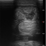
Case presentation - June 9, 2016: A 9 year old Warmblood jumping horse presented with a superficial digital flexor tendon injury of the right hind leg. The horse was treated with stem cell injections. After 10 months of rest and rehabilitation, the horse slowly went back to jumping but in late August 2018, the horse re-injured the SDFT. The horse underwent shockwave treatments and experienced marginal improvement. On January 18, 2019, after more than 2 years following the initial injury and a multitude of treatments, 1.5 cc of RenoVō® was implanted at the discretion of the veterinarian into the lesion under ultrasound guidance.


RenoVō® Case Study: Tendon Healing in a Competitive Warmblood Imaging 2
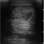
February 27, 2019: Only 1 Month after the RenoVō® implantation, the attending veterinarian observed a significant improvement under ultrasound. At the recommendation of the veterinarian, the horse continued resting.


RenoVō® Case Study: Tendon Healing in a Competitive Warmblood Imaging 3
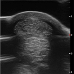
April 22, 2019: 13 Months following the RenoVō® implantation, the lesion was finally resolved, and horse was sound. "In October 2019, Hands Free went back to jumping and competed at 1.15m, jumping clean the entire week and had no change in his soundness," says the owner. The horse is still sound to date and is now showing at 1.25m.